


Welcome to MHP Imaging at Comprehensive Medical Center. We are here to provide you with the best quality care for your imaging needs. We are proud to offer CT scan, Ultrasound and X-ray.
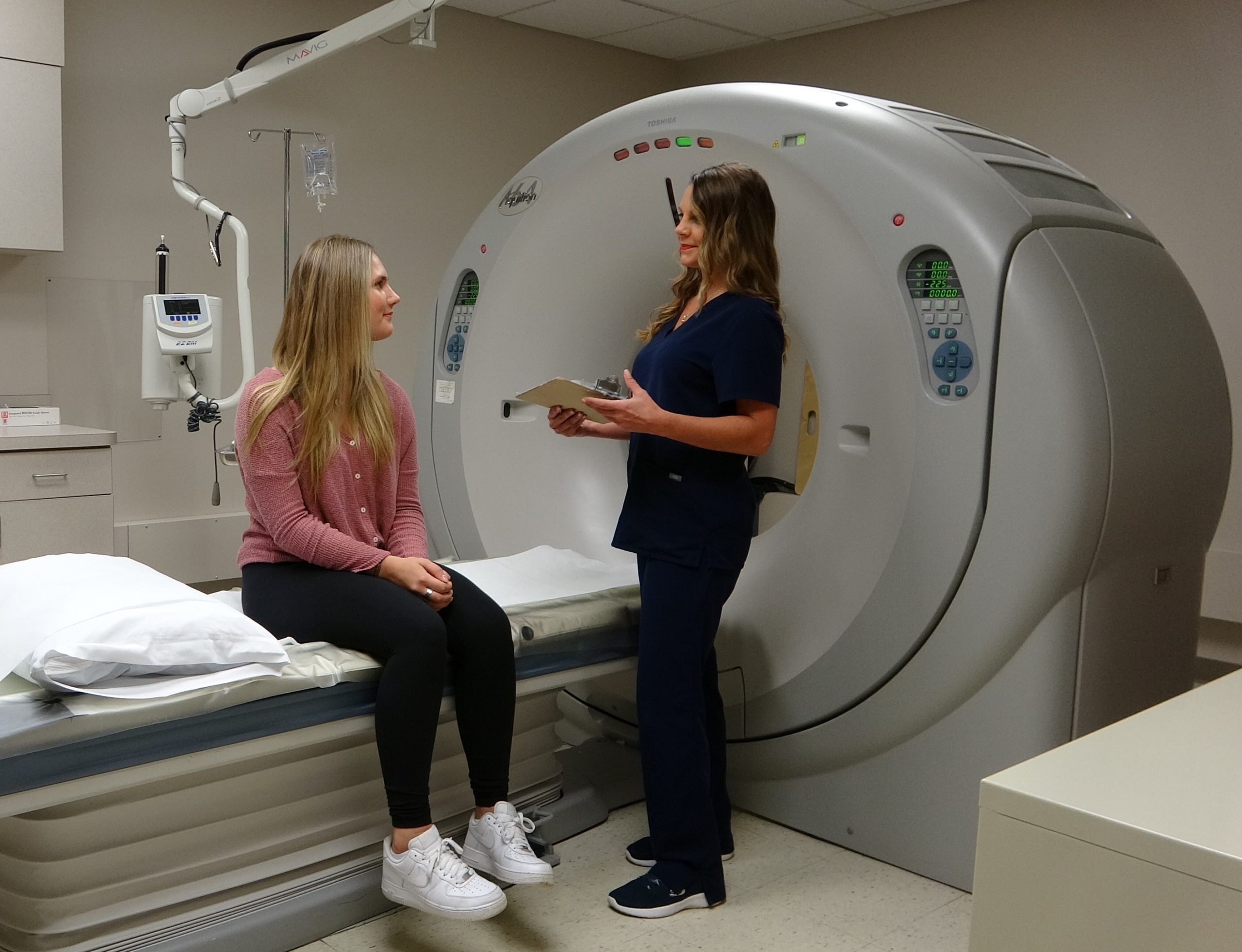
Our newly upgraded Toshiba Aquillion 64 slice is a state of the art CT machine. It is equipped with advanced software that features 40% dose reduction compared to similar systems while delivering the highest image quality to ensure accurate diagnosis . This scanner is one of the fastest computed tomography (CT) scanners on the market today, requiring less time to complete the exam. Our caring technologists and staff have your best interests in mind. They will spend time with you to explain your exam and make necessary preparations to ensure the best imaging possible.
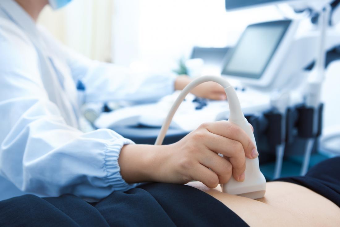
An ultrasound exam (or “sonogram”) is a painless diagnostic technique that makes use of how sound waves travel through the body. When sound waves pass through the body, they bounce off tissues and organs in certain ways. The reflected waves can be used to make images of the organs inside. The sound waves don’t hurt the body, and there’s no radiation
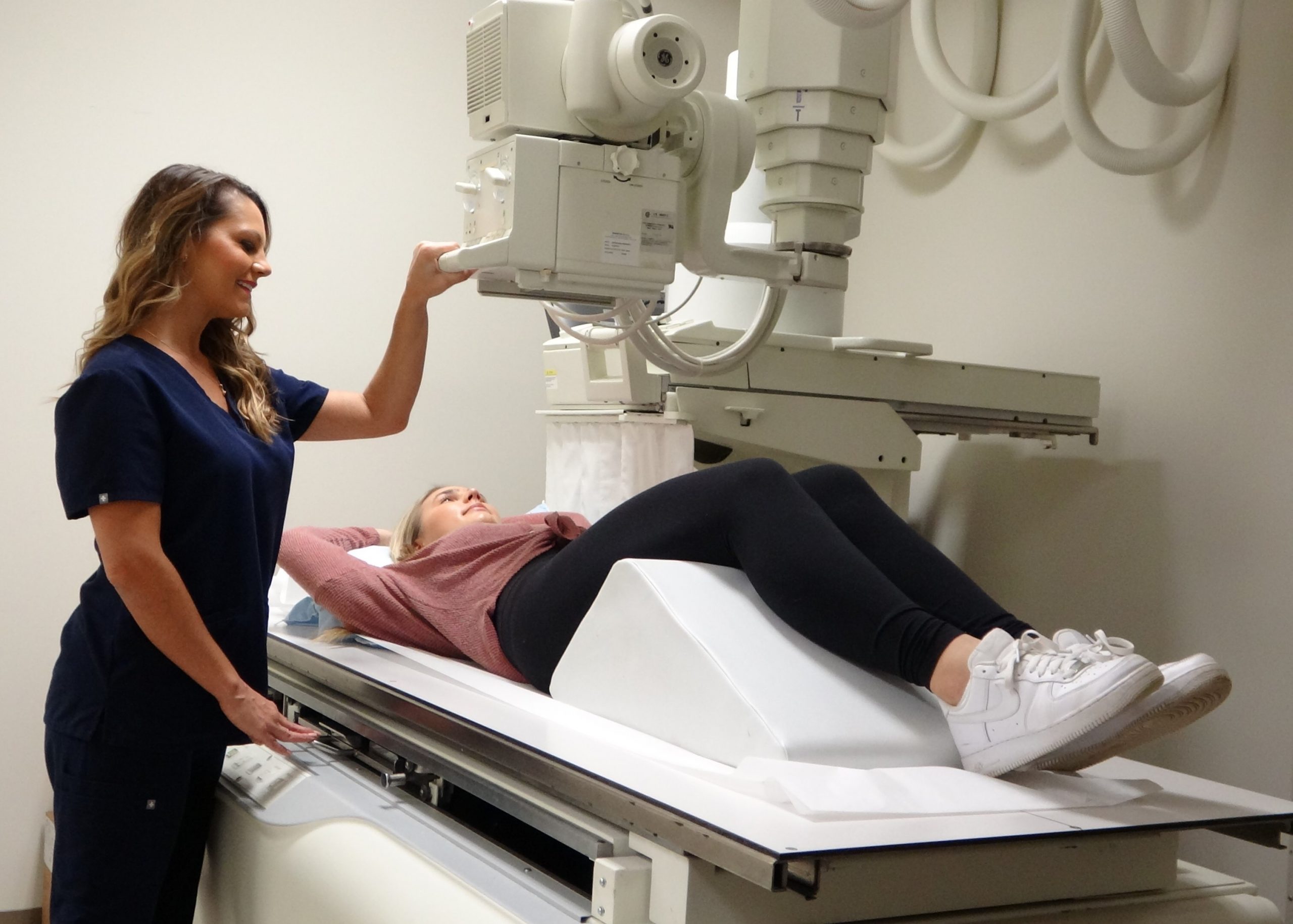
A kidney, ureter, and bladder (KUB) X-ray may be prescribed by your provider to assess the abdominal area for causes of abdominal pain, or to assess the organs and structures of the urinary and/or gastrointestinal (GI) system. A KUB X-ray may be the first diagnostic procedure used to assess the urinary system
Getting an accurate diagnosis can be one of the most impactful experiences that you can have — especially if you’ve been in search of that answer for a while. We can help you get there.
We are conveniently located at 31157 Woodward Ave in Royal Oak, on Woodward Ave just north of 13 Mile. We are open Monday- Friday 7:30 AM-4:00 PM. You will find our office to be comfortable and our staff to be friendly. Parking is convenient and our one-story building is easy and quick for you to navigate through. We look forward to serving you.
Prior to your CT scan we request that you do not eat anything 4 hours prior to your exam time. All regular medications can be taken before your exam.
If your CT scan has been ordered with oral contrast, you will drink your first bottle of contrast 2 hours prior to your exam. The 2nd bottle one hour prior to your exam.
Urogram studies require you to drink 40 oz of water prior to your exam. It is important to know that you are able to use the restroom as often as you need to.
You will be receiving a phone call from one of our staff members a day prior to your exam to go over your prep and answer any additional questions that you may have.
If your test has been ordered with IV contrast and you have not had a creatine level (blood draw) within 30 days our office has a POC I STAT machine and is able to obtain the lab value within less than 2 minutes. A blood sample will be drawn at the time your IV is started for your CT Exam.
On the day of the exam, wear comfortable, loose-fitting clothing. Metal objects can affect the image, so avoid clothing with zippers and snaps. You must put on a hospital gown and remove watches, jewelry, or any other metal objects you might be wearing (including hairpins, glasses, hearing aids and any removable dental work).
You will be asked to remove any metallic or other items and ALL jewelry that might interfere with the scan.
Your CT scan will take about 15 minutes. Certain types of examinations require that intravenous contrast (“x-ray dye”) be administered in order to best evaluate the organ system or disease. During the scan you will be lying on a padded table. You may be asked to lie on your stomach, back or side, and to hold your breath or stay very still. You may hear humming noises or feel the table move slowly through the CT scanner.
After your CT scan, if you received contrast, you should drink about five glasses of water to help flush out the contrast that was injected.
After you have completed the computed tomography (CT) scan, your health care team will review the images to make sure they are of an acceptable level of quality. If more images are needed, they will usually be taken right away. Usually your doctor will contact you with the CT results within 2 business days. If you don’t hear from your doctor after 4 or 5 business days of your exam please contact our imaging department @ at 248-336-3175.
Ultrasounds can be used to look at different organs in the body. Below is a list of ultrasounds that we perform in our office:
The bladder is an organ made of smooth muscle. It stores urine until it’s released when you go to the bathroom. The most common reason for bladder ultrasound is to check bladder draining. The urine that remains in the bladder after urinating (“post void residual”) is measured. If urine remains, there can be a problem like: Enlarged prostate, Urethral stricture (narrowing), Bladder dysfunction, Bladder ultrasound can also give information about:
The bladder wall, Diverticula (pouches) of the bladder, Prostate size, Stones, Large tumors in the bladder, Bladder ultrasound doesn’t check the ovaries, uterus, or colon.
This test doesn’t require fasting or bowel preparation. If you are not checking for post void residuals, a full bladder is needed. You may be asked to drink many glasses of water an hour before the exam.
The exam is done as you lay on your back on the exam table. A gel is spread on the skin to help transmit the sound waves. The transducer is placed between your navel and pubic bone. The image is viewed on a monitor and read on the spot. To check bladder draining, you’ll be asked to void. When you return, the bladder will be imaged again.
The kidneys are 2, fist-sized organs found on either side of your mid-section (“retroperitoneum”). The kidneys remove waste from the blood and make urine. They also balance salts (“electrolytes”) in the body, such as sodium and potassium. Hormones that control blood pressure and red blood cell production are also made in the kidneys.
Renal ultrasound studies can show the size and position of the kidneys. They can also show if there are: Blockages, Stones or Tumors.
A kidney ultrasound creates images from sound waves that return from the kidney tissue. Many images are collected to understand problems in the kidney.
If your doctor wants to see how blood flows to and from the kidney, Doppler imaging is used. This is an ultrasound method that makes color images from the movement of flowing blood. It shows the flow of blood through the vessels. It provides excellent motion information not available on a standard sonogram.
No need to fast, prepare your bowel, or have a full bladder. The test is done as you lay on your back on the exam table. A gel is spread on the skin to help transmit the sound waves. The kidneys are imaged by placing the transducer over both sides of the upper belly.
Ultrasound imaging of the scrotum uses sound waves to produce pictures of a male’s testicles and surrounding tissues. It is the primary method used to help evaluate disorders of the testicles, epididymis (tubes immediately next to the testicles that collect sperm) and scrotum. Ultrasound is safe, noninvasive, and does not use ionizing radiation. This procedure requires little to no special preparation. Leave jewelry at home and wear loose, comfortable clothing. You may be asked to wear a gown.
A prostate biopsy is a procedure to remove samples of suspicious tissue from the prostate. The prostate is a small, walnut-shaped gland in men that produces fluid that nourishes and transports sperm.
During a prostate biopsy a needle is used to collect a number of tissue samples from your prostate gland. The procedure is performed by a doctor who specializes in the urinary system and men’s sex organs (urologist). Your urologist will provide you with specific instruction to prepare for this test.
